
Approved by the Cancer.Net Editorial Board, 09/2023
ON THIS PAGE: You will find a drawing of the main body parts affected by cervical cancer. Use the menu to see other pages.
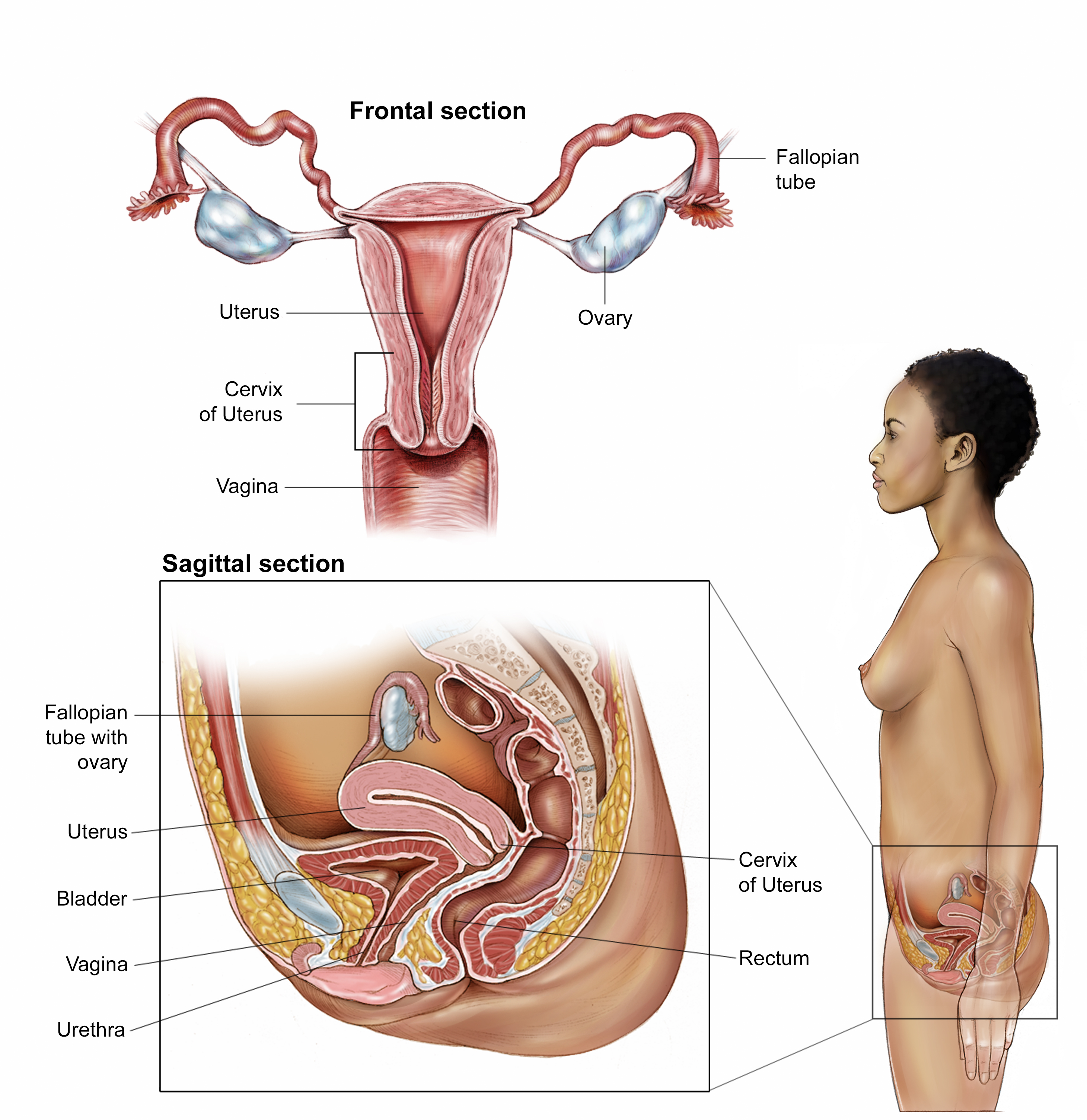
This illustration shows a frontal and sagittal (side) view of a woman’s reproductive system. The frontal section shows the fallopian tubes, 2 small ducts that link the 2 ovaries (1 on each side) to the hollow, pear-shaped uterus. The lower, narrow part of the uterus is called the cervix, which leads to the vagina. The uterus is located in the pelvis, between the bladder and rectum, and the vagina is located behind the urethra, which connects to the bladder. Copyright 2003 American Society of Clinical Oncology. Robert Morreale/Visual Explanations, LLC.
Copyright 2022 American Society of Clinical Oncology. Robert Morreale.
The next section in this guide is Risk Factors and Prevention. It describes the factors that may increase the chance of developing cervical cancer. Use the menu to choose a different section to read in this guide.