
ON THIS PAGE: You will find basic information about this group of diseases and the parts of the body they may affect. This is the first page of Cancer.Net’s Guide to Ovarian, Fallopian Tube, and Peritoneal Cancer. Use the menu to see other pages. Think of that menu as a roadmap for this entire guide.
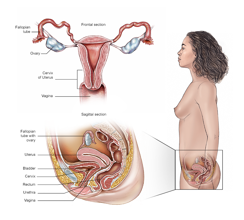
About the ovaries
The ovaries are part of the female reproductive system. There are 2 ovaries, with 1 located on each side of the uterus. Before menopause occurs, ovaries are almond-shaped and about 1.5 inches long. They contain eggs, also called germ cells. Ovaries are the primary source of estrogen and progesterone for the body. These hormones influence breast growth, body shape, body hair, and regulate the menstrual cycle and pregnancy. During and after menopause, the ovaries stop releasing eggs and producing certain hormones.
About the fallopian tubes
The fallopian tubes are small ducts that connect the ovaries to the uterus. They are part of the female reproductive system. Typically, the female reproductive system has 2 fallopian tubes, with 1 located on each side of the uterus. During ovulation, which typically happens monthly, an egg is usually released from 1 ovary and travels through a fallopian tube to the uterus.
About the peritoneum
The peritoneum is a tissue that lines the abdomen and most of the organs in the abdomen. The tissue covers the uterus, bladder, rectum, and the ovaries and fallopian tubes. A fold of this tissue called the omentum covers and connects the organs in the abdomen. A liquid called peritoneal fluid covers the tissue’s surface. This liquid helps the organs move within the abdomen and prevents them from sticking together.
About ovarian, fallopian tube, and peritoneal cancers
The term “ovarian cancer” is often used to describe cancers that begin in the cells in the ovary, fallopian tube, or peritoneum. The cancers are closely related and are treated the same way.
In this guide, this group of cancers is referred to as “ovarian/fallopian tube cancer” because peritoneal cancer is relatively rare. When the term “ovarian cancer” is used, it includes both fallopian tube and peritoneal cancers because it may be unclear where the cancer started.
These types of cancer begin when healthy cells in these areas change and grow out of control, forming a mass called a tumor. A tumor can be cancerous or benign. A cancerous tumor is malignant, meaning it can grow, invade, and spread to other parts of the body. A benign tumor means the tumor can grow but will not spread.
An ovarian cyst is an abnormal growth of tissue that forms on the surface of the ovary and includes fluid. It can occur during a normal menstrual cycle and usually goes away without treatment. Simple ovarian cysts are not cancerous.
Recent research studies suggest that most ovarian/fallopian tube/peritoneal cancers are high-grade serous cancers (HGSC; see more information below), and in most cases, the cancer actually starts in the tip, or outer end, of the fallopian tubes. Then, it spreads to the surface of the ovaries and can spread beyond.
Based on this updated knowledge, when discussing contraception to avoid future pregnancy, some doctors recommend removal of the fallopian tubes, rather than tying or banding the tubes, in order to lower the risk of ovarian/fallopian tube cancers. Some doctors also recommend fallopian tube removal when a person is undergoing surgery for a benign disease and does not want to get pregnant in the future. This strategy could reduce the chance of these cancers developing in the future. Learn more about prevention in another section of this guide.
Because the surfaces of the ovaries, the lining of the fallopian tubes, and the covering cells of the peritoneum are made up of the same types of cells, most of these diseases look alike under a microscope. Rarely, peritoneal cancer can develop after ovaries and fallopian tubes have been removed. Just as with ovarian cancer, some peritoneal cancers may begin in the fallopian tubes and spread from the end of the fallopian tube into the peritoneal cavity.
Types of ovarian and fallopian tube cancer
-
Epithelial carcinoma. Epithelial carcinoma makes up 85% to 90% of ovarian/fallopian tube cancers. The main types of epithelial tumors include serous, endometrioid, clear cell, mucinous, mixed tumors, and several rare cancers, including Brenner tumors. These types describe how these different ovarian/fallopian tube cancers are classified based on how they look under the microscope. There can be differences in how these cancers behave and which treatments will work best.
The vast majority of epithelial cancers are HGSC, meaning they resemble the cells lining the fallopian tube. HGSCs make up the vast majority of ovarian/fallopian tube cancer, most of which arise from the fallopian tube. LGSC is less common and may arise from the ovaries.
-
Germ cell malignancies. This rare type of ovarian cancer develops in the egg-producing cells of the ovaries. Germ cell malignancies typically occur between the ages of 10 to 29.
Types of germ cell tumors include dysgerminomas, immature teratoma, endodermal sinus tumors (called EST and yolk sac tumors), and embryonal carcinomas.
-
Sex cord stromal tumors. This rare form of ovarian tumor develops in the connective tissue cells, called granulosa and theca cells, that hold the ovaries together. This tissue sometimes makes the female hormones estrogen and progesterone. Over 90% of these stromal tumors are called granulosa cell tumors, either adult or juvenile types.
Granulosa cell tumors may secrete estrogen, resulting in unusual vaginal bleeding at the time of diagnosis. Other types are Sertoli-Leydig cell tumors and steroid cell tumors.
-
Fallopian tube cancer. This cancer was once thought to be rare, but science now shows that most cancers previously labeled “ovarian cancer” actually begin in a fallopian tube. Virtually all of these are serous cancers, and most are HGSC. However, in rare cases, other types of cancer can start in the fallopian tube.
Looking for More of an Introduction?
If you would like more of an introduction, explore these related items. Please note that these links will take you to other sections on Cancer.Net:
-
ASCO Answers Fact Sheet: Read a 1-page fact sheet on ovarian/fallopian tube cancer that offers an introduction. This free fact sheet is available as a PDF so it is easy to print.
-
Cancer.Net En Español: Read about ovarian, fallopian tube, and peritoneal cancer in Spanish. Infórmase sobre cáncer de ovario, fallopian tube, peritoneum en español.
-
Find a Cancer Doctor. Search for a cancer specialist in your local area using this free database of doctors from the American Society of Clinical Oncology (ASCO).
-
Cancer Terms. Learn what medical phrases and terms used in cancer care and treatment mean.
The next section in this guide is Statistics. It helps explain the number of people who are diagnosed with ovarian, fallopian tube, and peritoneal cancer and the general survival rates. Use the menu to choose a different section to read in this guide.