
Approved by the Cancer.Net Editorial Board, 03/2023
ON THIS PAGE: You will find drawings of the main body parts affected by a brain tumor. Use the menu to see other pages.
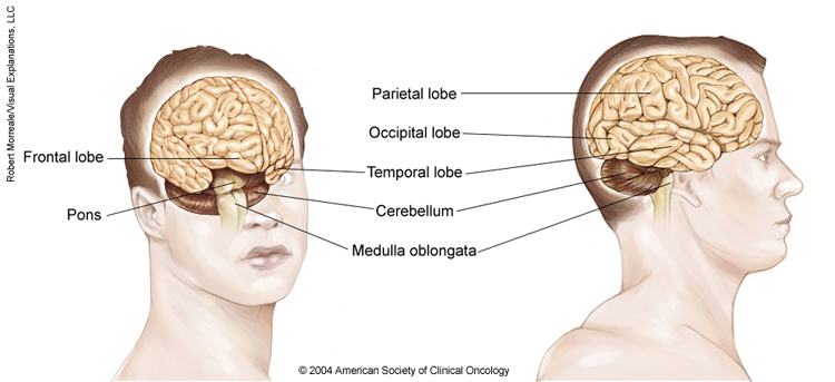
The image is two drawings showing different parts of a person’s brain. The largest part of the brain, the cerebrum, is made up of four lobes: the frontal lobe at the front of the skull, the parietal lobe at the upper rear of the skull, above the occipital lobe, which is at the back of the skull, and the temporal lobe, which is located under the frontal and parietal lobes on both sides of the cerebrum. The cerebellum is located under the occipital and temporal lobes at the rear of the skull. The brain stem includes the pons and the medulla oblongata, which connects to the spinal cord. Copyright 2004 American Society of Clinical Oncology. Robert Morreale/Visual Explanations, LLC.
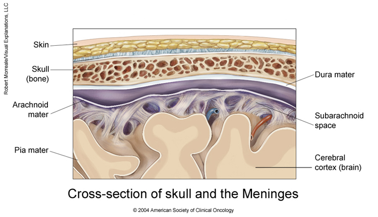
The image is a drawing of a cross-section of the skull and meninges. It shows the layers of membranes that surround and protect the brain. The first layer shows the skin covering the skull bone. Beneath the skull is the dura mater, which surrounds the arachnoid mater. Cerebrospinal fluid circulates in the sub arachnoid space between arachnoid mater and the pia mater, which covers the cerebral cortex (the brain). Copyright 2004 American Society of Clinical Oncology. Robert Morreale/Visual Explanations, LLC.
The next section in this guide is Statistics. It helps explain the number of people who are diagnosed with a brain tumor and general survival rates. Use the menu to choose a different section to read in this guide.