
ON THIS PAGE: You will learn about how doctors describe a cancer’s growth or spread, as well as what the cancer cells look like under a microscope. This is called the stage and grade. Use the menu to see other pages.
Staging is a way of describing where the cancer is located, if or where it has spread, and whether it is affecting other parts of the body.
Doctors use diagnostic tests to find out the cancer’s stage, so staging may not be complete until the doctor performs a biopsy. Knowing the stage helps the doctor to decide what kind of treatment is best and can help predict a person's prognosis, which is the chance of recovery. There are different stage descriptions for different types of cancer.
TNM staging system
One tool that doctors use to describe the stage is the TNM system. Doctors use the results from diagnostic tests and scans to answer these questions:
-
Tumor (T): How large is the primary tumor? Where is it located?
-
Node (N): Has the tumor spread to the lymph nodes? If so, where and how many?
-
Metastasis (M): Has the cancer spread to other parts of the body? If so, where and how much?
The results are combined to determine the stage of cancer for each person. There are 5 stages: stage 0 (zero) and stages I through IV (1 through 4). The stage provides a common way of describing the cancer, so doctors can work together to plan the best treatments.
Staging can be clinical or pathological. "Clinical staging" is based on the results of tests done before surgery, which may include physical examinations and imaging tests. "Pathological staging" is based on what is found during surgery. In general, pathological staging provides the most information to determine a patient’s prognosis.
Here are more details on each part of the TNM system for salivary gland cancer:
Tumor (T)
Using the TNM system, the "T" plus a letter or number (0 to 4) is used to describe the size and location of the tumor. Tumor size is measured in centimeters (cm). A centimeter is roughly equal to the width of a standard pen or pencil.
Stage may also be divided into smaller groups that help describe the tumor in even more detail. Specific tumor stage information is listed below.
TX: Indicates the primary tumor cannot be evaluated.
T0 (T plus zero): No evidence of a tumor was found.
T1: Describes a small, noninvasive (has not spread) tumor that is 2 cm at its greatest dimension.
T2: Describes a larger, noninvasive tumor between 2 cm to 4 cm.
T3: Describes a tumor that is larger than 4 cm but not larger than 6 cm and has spread beyond the salivary glands but does not affect the seventh nerve, the facial nerve that controls expression, such as smiles or frowns.
T4a: The tumor has invaded the skin, jawbone, ear canal, and/or facial nerve.
T4b: The tumor has invaded the skull base and/or the nearby bones and/or encases the arteries.
Node (N)
The “N” in the TNM staging system is for lymph nodes. Lymph nodes near the head and neck are called regional lymph nodes. Lymph nodes in other parts of the body are called distant lymph nodes. There are many nodes located in the head and neck area, and careful evaluation of lymph nodes is an important part of staging cancer of the major salivary glands.
NX: The regional lymph nodes cannot be evaluated.
N0: There is no evidence of cancer in the regional lymph nodes.
N1: Cancer has spread to a single lymph node on the same side as the primary tumor, and the cancer found in the lymph node is 3 cm or smaller.
N2: Describes any of these conditions:
- N2a: The cancer has spread to a single lymph node on the same side as the primary tumor and is larger than 3 cm but smaller than 6 cm.
- N2b: The cancer has spread to more than 1 lymph node on the same side as the primary tumor, and none measures larger than 6 cm.
- N2c: The cancer has spread to more than 1 lymph node on either side of the body, and none measures larger than 6 cm.
N3: The cancer found in the lymph nodes is larger than 6 cm.
Metastasis (M)
The "M" in the TNM system describes cancer that has spread to other parts of the body, called distant metastasis.
MX: Indicates distant metastasis cannot be evaluated.
M0: Indicates the cancer has not spread to other parts of the body.
M1: Describes cancer that has spread to other parts of the body.
Cancer stage grouping
Doctors assign the stage of the cancer by combining the T, N, and M classifications.
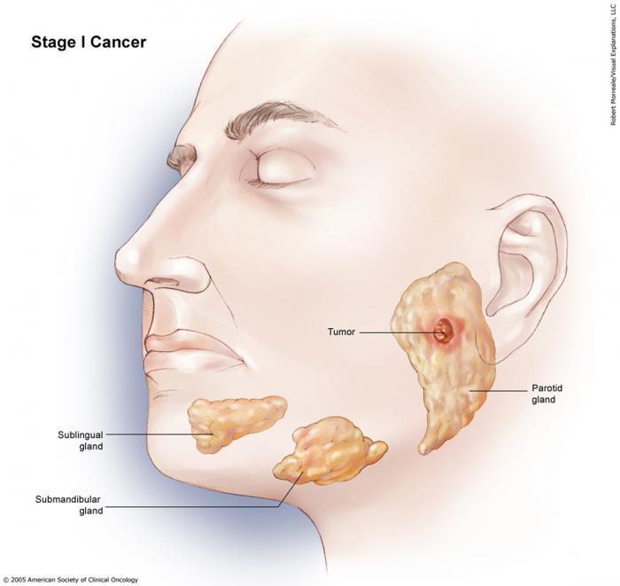
Stage I: Noninvasive tumors (T1, T2) with no spread to lymph nodes (N0) and no distant metastasis (M0).
Stage II: An invasive tumor (T3) with no spread to lymph nodes (N0) or distant metastasis (M0).

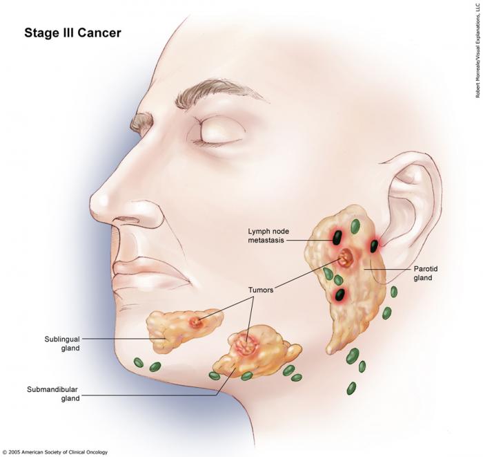
Stage III: Smaller tumors (T1, T2) that have spread to regional lymph nodes (N1) but have no sign of metastasis (M0).
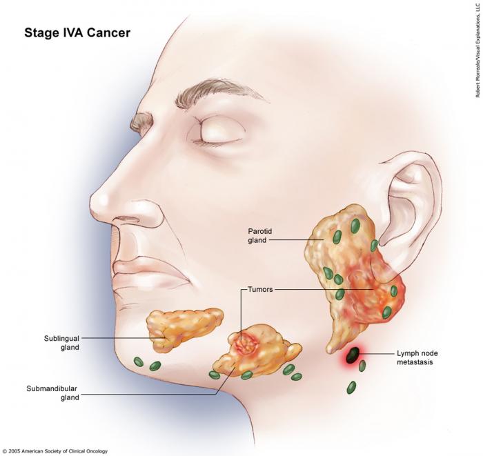
Stage IVA: Any invasive tumor (T4a) with either no lymph node involvement (N0) or spread to only a single same-sided lymph node (N1), but no metastasis (M0). It is also used for a T3 tumor with 1-sided lymph nodal involvement (N1) but no metastasis (M0), or any tumor (any T) with extensive lymph nodal involvement (N2).
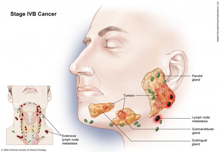
Stage IVB: Any cancer (any T), with more extensive spread to lymph nodes (N2, N3), but no metastasis (M0).
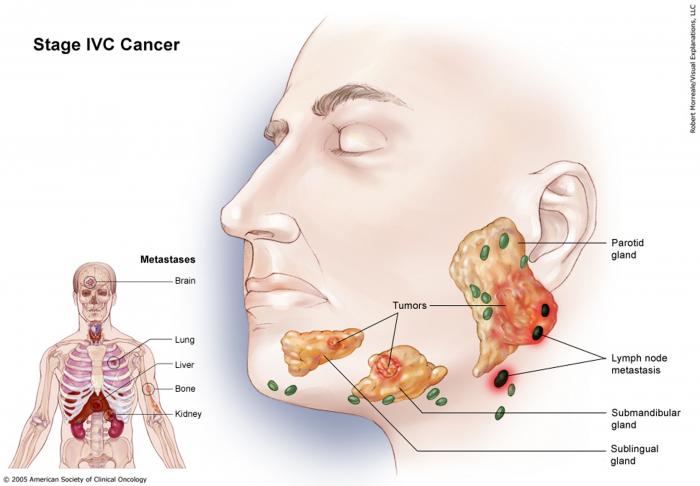
Stage IVC: Any cancer with distant metastasis (any T, any N, and M1).
Recurrent: Recurrent cancer is cancer that has come back after treatment. If the cancer does return, there will be another round of tests to learn about the extent of the recurrence. These tests and scans are often similar to those done at the time of the original diagnosis.
Used with permission of the American College of Surgeons, Chicago, Illinois. The original and primary source for this information is the AJCC Cancer Staging Manual, Eighth Edition (2017) published by Springer International Publishing.
Grade (G)
Doctors also describe this type of cancer by its grade (G). The grade describes how much cancer cells look like healthy cells when viewed under a microscope.
The doctor compares the cancerous tissue with healthy tissue. Healthy tissue usually contains many different types of cells grouped together. If the cancer looks similar to healthy tissue and has different cell groupings, it is called “differentiated” or a “low-grade tumor.” If the cancerous tissue looks very different from healthy tissue, it is called “poorly differentiated” or a “high-grade tumor.” The cancer’s grade may help the doctor predict how quickly the cancer will spread. In general, the lower the tumor’s grade, the better the prognosis.
Information about the cancer’s stage and grade will help the doctor recommend a specific treatment plan. The next section in this guide is Types of Treatment. Use the menu to choose a different section to read in this guide.